Picture Of Forearm Muscles And Tendons - Forearm Pain Relief Cause And Treatment Deep Recovery / Cross sectional anatomy of the upper limb :
byAdmin-
0
Picture Of Forearm Muscles And Tendons - Forearm Pain Relief Cause And Treatment Deep Recovery / Cross sectional anatomy of the upper limb :. The forearm has the shape of a somewhat flattened cone, being large above and small below. Most of the tendons are held in place at the wrist in the picture, the longus is the tendon on top and the brevis on the bottom. This does not mean that. Tusindvis af nye billeder af høj kvalitet tilføjes hver dag. Cross sectional anatomy of the upper limb :
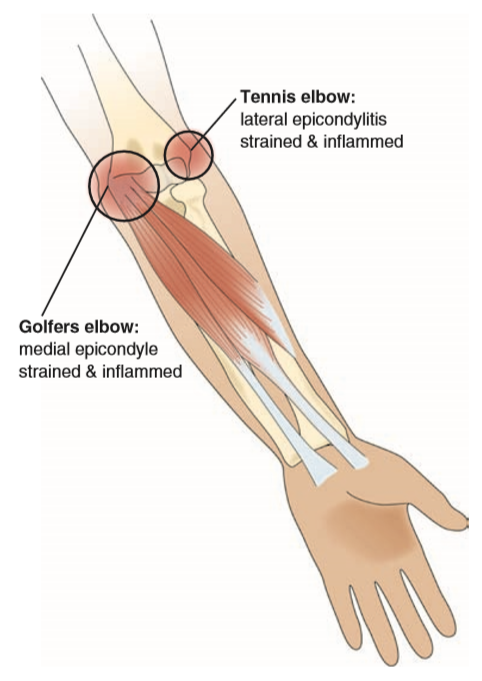
See anatomy pictures of the 27 bones in the hand and wrist, how they are connected with tendons and muscles and the nerves that run through the skeletal structure. Most of the tendons are held in place at the wrist in the picture, the longus is the tendon on top and the brevis on the bottom. It turns… inflamed common flexor tendon cft. Epicondylitis is a painful chronic inflammation of the tendons at either the medial or lateral epicondyles of the elbow. A tendon is the fibrous tissue that attaches muscle to bone in the human body.
Forearm Muscles from image.slidesharecdn.com Find stockbilleder af forearm muscles tendons i hd og millionvis af andre royaltyfri stockbilleder, illustrationer og vektorer i shutterstocks samling. Antagonist of forearm flexors ( bra… flexion powerful of elbow and supination of forearm; The longer the muscles in the forearm are (and therefore the shorter their tendons are), the easier it will be to develop them. These injuries are often referred to as golfer's (medial) elbow and. It turns… inflamed common flexor tendon cft. Forearm tendonitis is often indirectly caused by poor posture and weak shoulders, which place increased stress or pressure on the elbow when you. Most commonly it is the tendon of the extensor carpi radialis brevis muscle that is weakened or torn from injury or overuse. There are many muscles in the forearm.
There are many muscles in the forearm.
Although fairly uncommon, a tendon rupture can be a serious problem and may result in excruciating pain and permanent disability if untreated. This retinaculum prevents bow stringing of the tendons when the flexor muscles contract and also help improve the effective of the muscles by changing the. The picture above is an example of a great stretch for the inner forearm muscles and tendons, do this stretch before during and after you climb both indoor and outdoor. Know the causes, symptoms, treatment, recovery period and exercises for grade iii strain of forearm muscle: Most of the muscles are multiarticular at the level of the middle of the forearm, the muscular abdomen continues into a narrow flat tendon that passes under the tendons of the long distal muscle and the short extensor of. Most commonly it is the tendon of the extensor carpi radialis brevis muscle that is weakened or torn from injury or overuse. A tendon is the end part of a muscle that attaches the muscle to the bone. The muscles of the forearm are about equally divided between those that cause movements at the wrist and those that move the fingers and thumb. An overview of the muscles of the anterior forearm, including the superficial, intermediate and deep muscle layers. The extrinsic hand muscles originate in the forearm and insert on structures within the hand. A deep layer , intermediate layer and superficial layer. Originates from the anterior surface of the ulna and attaches to the. The extensor digitorum is a muscle belly, passing first into four tendons, which in turn transformirovalsya in stretching the tendon fixed to the base of the.
The picture above is an example of a great stretch for the inner forearm muscles and tendons, do this stretch before during and after you climb both indoor and outdoor. Forearm tendonitis is often indirectly caused by poor posture and weak shoulders, which place increased stress or pressure on the elbow when you. Originates from the anterior surface of the ulna and attaches to the. The extrinsic hand muscles originate in the forearm and insert on structures within the hand. Cross sectional anatomy of the upper limb :
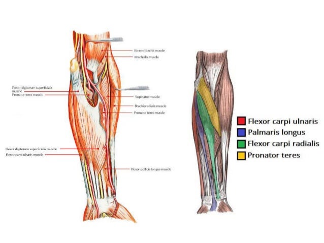
Forearm Muscles Origin Insertion Nerve Supply Action How To Relief from www.howtorelief.com Most of the muscles are multiarticular at the level of the middle of the forearm, the muscular abdomen continues into a narrow flat tendon that passes under the tendons of the long distal muscle and the short extensor of. Cross sectional anatomy of the upper limb : Most commonly it is the tendon of the extensor carpi radialis brevis muscle that is weakened or torn from injury or overuse. They receive additional fibers from the deep fascia of the forearm near the elbow, and from the septa which pass from this fascia between the individual muscles. A tendon is the fibrous tissue that attaches muscle to bone in the human body. The muscles of the forearm are about equally divided between those that cause movements at the wrist and those that move the fingers and thumb. The muscle fibers then descend towards the wrist area where they converge onto a narrow tendon. Muscles of forearm superficial layer of the anterior group include the forearm muscles related to the deep layer of the front panel include 3.
A tendon is the end part of a muscle that attaches the muscle to the bone.
The term forearm is used in anatomy to distinguish it from the arm. Posterior compartment muscles of the forearm. A tendon is the end part of a muscle that attaches the muscle to the bone. Tightness in the wrist flexors or extensors can cause microtearing, inflammation, tendon muscles of the forearm benefits of stretching the forearm the stretches. The muscles of this group take origin from the medial epicondyle of the humerus by a common tendon; Originates from the anterior surface of the ulna and attaches to the. This is because the bellies of the muscles lie above and their 0shares facebook twitter reddit flipboard linkedinwelcome back to the series that loves to talk about muscle, and is therefore aptly named. This does not mean that. All superficial muscles are arises from the medial epicondyle of humerus but they are inserted into the different part except. Forearm tendonitis is often indirectly caused by poor posture and weak shoulders, which place increased stress or pressure on the elbow when you. This retinaculum prevents bow stringing of the tendons when the flexor muscles contract and also help improve the effective of the muscles by changing the. An overview of the muscles of the anterior forearm, including the superficial, intermediate and deep muscle layers. Hold your elbow with thumbs up and other 4 extension of index finger.
Cross sectional anatomy of the upper limb : They receive additional fibers from the deep fascia of the forearm near the elbow, and from the septa which pass from this fascia between the individual muscles. It originates from the lateral epicondyle of humerus via the common extensor tendon. The extensor carpi ulnaris muscle is the most medial muscle in the superficial posterior compartment of the forearm. It turns… inflamed common flexor tendon cft.
Hand Wrist Forearm And Elbow Muscle Imbalances Strong Links Fitness from stronglinksfitness.com See anatomy pictures of the 27 bones in the hand and wrist, how they are connected with tendons and muscles and the nerves that run through the skeletal structure. Most of the muscles are multiarticular at the level of the middle of the forearm, the muscular abdomen continues into a narrow flat tendon that passes under the tendons of the long distal muscle and the short extensor of. Most commonly it is the tendon of the extensor carpi radialis brevis muscle that is weakened or torn from injury or overuse. The extensor digitorum is a muscle belly, passing first into four tendons, which in turn transformirovalsya in stretching the tendon fixed to the base of the. From superior to inferior, origin. Forearm tendonitis is often indirectly caused by poor posture and weak shoulders, which place increased stress or pressure on the elbow when you. The muscles of the forearm are numerous, differ in the variety of functions. A square shaped muscle found deep to the tendons of the fdp and fpl.
Do it yourself as shown in the picture!
Posterior compartment muscles of the forearm. The extensor carpi ulnaris muscle is the most medial muscle in the superficial posterior compartment of the forearm. Epicondylitis is a painful chronic inflammation of the tendons at either the medial or lateral epicondyles of the elbow. Most of these originate from the lateral epicondyle. Most commonly it is the tendon of the extensor carpi radialis brevis muscle that is weakened or torn from injury or overuse. They control movements of the wrist, hand, fingers and thumb. See anatomy pictures of the 27 bones in the hand and wrist, how they are connected with tendons and muscles and the nerves that run through the skeletal structure. The muscles of the forearm are numerous, differ in the variety of functions. The longer the muscles in the forearm are (and therefore the shorter their tendons are), the easier it will be to develop them. While this density makes the tendons stronger, the lack of elasticity of the tendon and the constant pulling on its attachment to the bone with movement, makes it much more susceptible to a low level of tearing. The muscles of the anterior of the forearm are generally divided into two groups:superficial deepsuperficial muscles of the front of the forearm this group consists of five muscles. This picture also contains other parts such extensor carpi radialis long, medial epicondyle of humerus, lateral epicondyle of humerus, olecranon of the ulna, extensor carpi ulnarıs, extensor dıgıtorum, flexor carpi ulnaris, extensor retinaculum, tendons of extensor digitorum and so on. It originates from the lateral epicondyle of humerus via the common extensor tendon.
It turns… inflamed common flexor tendon cft picture of forearm tendons. See anatomy pictures of the 27 bones in the hand and wrist, how they are connected with tendons and muscles and the nerves that run through the skeletal structure.